Amoeba
Shape-shifting unicellular organisms found across all eukaryotic lineages.

Busition · CC BY 4.0
An amoeba (or ameba) is a type of cell or unicellular organism capable of altering its shape by extending and retracting pseudopods. Amoebae are not a single taxonomic group but are found in every major lineage of eukaryotic organisms, including protozoa, fungi, algae, and animals. They are often called amoeboid and are studied for their movement, feeding, and ecological roles.
- field
- Biology, Microbiology, Protistology
- known_for
- Amoeboid movement via pseudopods; phagocytosis; includes pathogens such as Entamoeba histolytica and Naegleria fowleri
- type
- Unicellular organism or cell type
- habitat
- Freshwater, marine, soil, and as parasites in hosts
Lore & Background
G. Ehrenberg.
Reader's Guide
Amoebae are significant as model organisms for studying cell movement, phagocytosis, and evolution. The best known amoeboid protists include Chaos carolinense and Amoeba proteus, widely used in classrooms. Pathogenic species such as Entamoeba histolytica (causing amoebic dysentery) and Naegleria fowleri (the 'brain-eating amoeba') have major health impacts. Recent evidence of meiosis-related genes in Acanthamoeba and Entamoeba suggests that sexual reproduction may be ancient in amoeboid lineages. Amoebae also serve as hosts for bacterial pathogens like Legionella, influencing disease spread. Their ecological roles range from predators to detritivores, and their size varies from 2.3 micrometres to 20 cm in xenophyophores.
Did You Know?
- Amoebae do not have cell walls, allowing free movement by extending pseudopods.
- The marine amoeboid Massisteria voersi is just 2.3 to 3 micrometres in diameter, similar to many bacteria.
- Some amoebae, such as Acanthamoeba and Dictyostelium discoideum, host bacterial pathogens like Legionella.
- The shells of deep-sea xenophyophores can reach 20 cm in diameter.
Taxonomic Diversity and the Collapse of Sarcodina
Amoebae defy the intuition that they belong to a single biological family. In reality, amoeboid cells appear across every major lineage of eukaryotic life, showing up not only among protozoa but also within fungi, algae, and animals. For much of the twentieth century, taxonomists lumped most of these shape-shifting organisms into a class or subphylum called Sarcodina, defined by the shared trait of moving through pseudopods or protoplasmic flow. That convenience, however, turned out to be a taxonomic illusion. Molecular phylogenetic studies demonstrated that Sarcodina is not monophyletic; its members do not all descend from a single common ancestor. As a result, modern classification no longer groups amoeboid organisms under one umbrella. Microbiologists today often use the terms amoeba and amoeboid interchangeably as functional descriptors for any organism that exhibits this characteristic mode of locomotion, rather than as markers of a specific evolutionary clade. The implication is profound: the ability to flow and extend cytoplasmic bulges either evolved independently multiple times or was inherited from a very ancient common ancestor, spanning the full breadth of eukaryotic diversity.
Pseudopods and the Mechanics of Amoeboid Movement
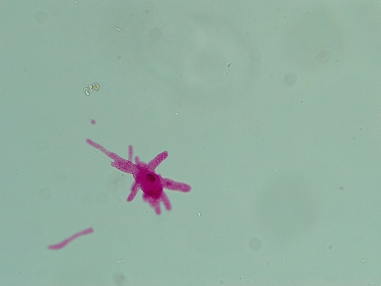
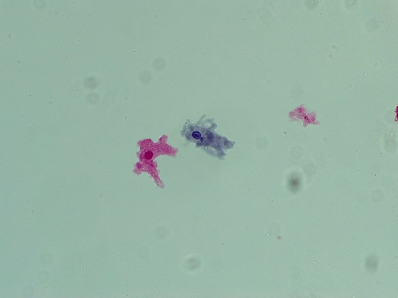
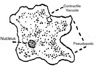
The hallmark of amoeboid life is the pseudopod, a temporary bulge of cytoplasm generated when actin microfilaments push the plasma membrane outward in a coordinated fashion. Because amoebae lack rigid cell walls, this membrane extension is free to reshape the entire body. The morphology of these extensions serves as a key diagnostic feature separating one amoeboid group from another. Amoebozoan species in the genus Amoeba produce bulbous, rounded lobose pseudopods that are roughly tubular in cross-section. Cercozoan forms such as Euglypha and Gromia extend slender, thread-like filose pseudopods. Foraminifera push out fine, branching pseudopods that interweave into net-like reticulose structures. At the opposite extreme, Radiolaria and Heliozoa project stiff, needle-like axopodia supported internally by bundles of microtubules. Beyond locomotion, some free-living amoebae build protective shells—testate forms—made of calcium, silica, chitin, or even agglutinated sand grains and diatom frustules, while others remain entirely naked with no hard covering whatsoever.
Feeding Strategies and Osmotic Survival
Amoebae are remarkably versatile feeders. Predatory species hunt bacteria and other protists, while detritivore species scavenge dead organic matter. The primary ingestion method is phagocytosis: pseudopods extend, encircle a particle or live prey, and engulf it into the cell. Unlike organisms with a fixed mouth or cytostome, amoeboid cells can perform phagocytosis at any point on their surface, with no predetermined feeding location. Some species supplement this with pinocytosis, drawing dissolved nutrients across the membrane through small internal vesicles. Osmotic regulation presents a separate survival challenge. Freshwater amoebae inhabit a hypotonic environment where the solute concentration outside the cell is lower than inside, causing water to flood in by osmosis. To prevent swelling and eventual rupture, most freshwater species maintain a contractile vacuole that actively pumps excess water out of the cytosol. Marine amoebae, by contrast, generally lack this organelle because the surrounding seawater is isotonic with their internal fluids, eliminating the constant osmotic influx of water.
Meiosis and the Ancient Sexuality of Amoebae
Long considered asexual, amoebae are now understood to harbor deep roots of sexual reproduction. Genomic analyses of Acanthamoeba revealed orthologs of key meiotic genes—Spo11, Mre11, Rad50, Rad51, Rad52, Mnd1, Dmc1, Msh, and Mlh—strongly suggesting that this lineage is capable of meiosis and, by extension, some form of sexual cycle. In Entamoeba histolytica, the meiosis-specific recombinase Dmc1 is actively expressed; purified Dmc1 forms presynaptic filaments and catalyses ATP-dependent homologous DNA pairing and strand exchange over thousands of base pairs, a reaction enhanced by the Hop2-Mnd1 heterodimer. Similar meiotic signatures appear in Entamoeba invadens, where recombination-related gene expression rises during encystation. Dictyostelium discoideum, a social amoeba, undergoes mating and meiosis when food becomes scarce. Because Amoebozoa diverged early in the eukaryotic tree, these findings point to meiosis as an ancient feature of eukaryotic evolution, supporting the hypothesis that most amoeboid lineages are anciently sexual.
Gallery



More in Microbiology 1-24
Elsewhere in the Microbiology universe
Spotted an error? Know more?
This is a living reference — every entry is fact-audited, and reader corrections feed straight into our audit queue. Suggest an edit · See this site's audit record