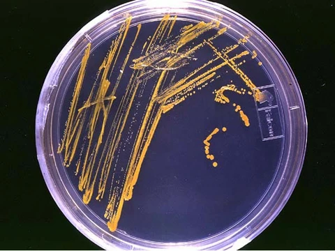
Endospore staining
A differential stain using heat to reveal bacterial endospores.

WMrapids · CC0
Endospore staining is a technique used in bacteriology to identify the presence of endospores in a bacterial sample. Endospores are protective structures formed by some bacteria to survive extreme conditions, including high temperatures, and they contain a tough outer coating of keratin. Because endospores are difficult to stain using normal techniques such as simple staining and gram staining, special methods like the Schaeffer–Fulton stain and the Moeller stain were developed.
- scientists_involved
- Cohn and Koch
- key_methods
- Schaeffer–Fulton stain, Moeller stain, Dorner method
- primary_stain
- Malachite green
- counterstain
- Safranin
Lore & Background
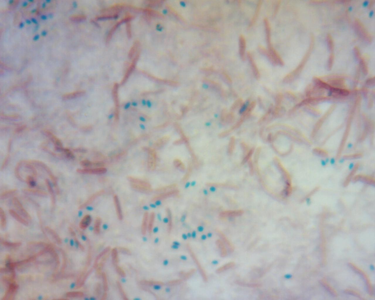
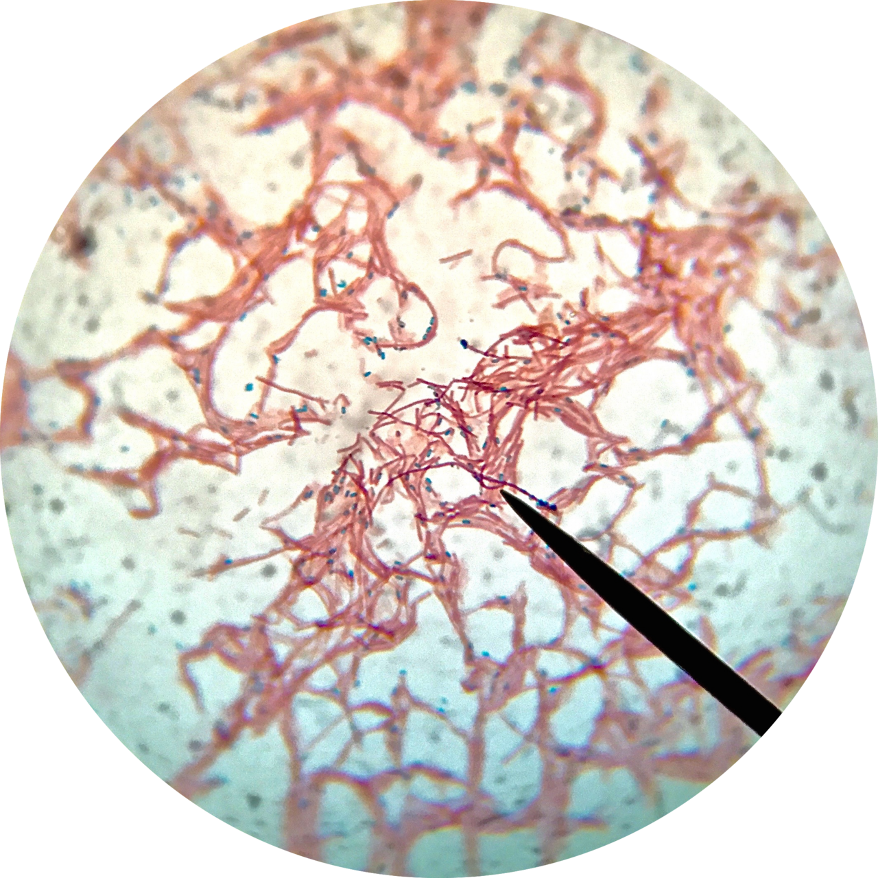
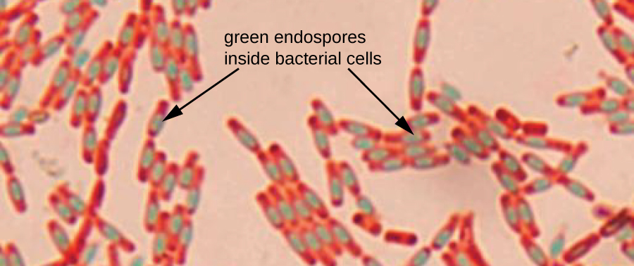
Although not the most beneficial, their method was more convenient and provided a quicker, easier test, making spores more susceptible to dyes. To this day, the Schaeffer-Fulton stain is still performed to help identify bacteria. The staining mechanism involves forcing malachite green into the spore by steaming the bacteria; heat acts as a mordant. Vegetative cells are decolorized with distilled water and counterstained with safranin, resulting in endospores appearing as green dots within red or pink cells.
Reader's Guide
Endospore staining is significant because it allows for the identification of bacteria that form endospores, such as Bacillus and Clostridium species, which include pathogens like Bacillus anthracis (causes anthrax), Clostridium tetani (causes tetanus), and Clostridium botulinum (found in improperly canned foods). The technique overcomes the resistance of endospores to standard staining methods. The Schaeffer-Fulton method, a refinement of Dorner's original work, remains a standard laboratory procedure. However, Mycobacterium can be an obstacle, as its waxy cell wall retains malachite green even after decolorization, requiring an acid-fast stain for further identification. Endospores can last for decades in harsh conditions like drying and freezing, and their shape and location (central, subterminal, terminal, free) aid in bacterial identification.
Did You Know?
- Endospores contain little or no ATP, indicating how dormant they can be.
- The Schaeffer-Fulton stain uses a Bunsen burner to speed up the heating process compared to Dorner's original method.
- Mycobacterium can falsely stain green in endospore staining due to its waxy cell wall.
- Endospores can be spherical or elliptical and may cause the cell to appear swollen.
The Staining Challenge and Differential Techniques
Visualizing endospores under a light microscope presents a genuine difficulty for the microbiologist, because the endospore wall is essentially impermeable to the dyes and stains that readily color the surrounding vegetative cell. The result is a frustrating contrast: the rest of the bacterial body takes up stain beautifully, while the spore sits there stubbornly colorless and nearly invisible. To overcome this barrier, dedicated staining protocols were devised. The Moeller stain is one such method, which renders the endospore a vivid red while the remainder of the cell is counterstained blue. Another widely employed approach is the Schaeffer-Fulton stain, which produces the reverse visual effect: endospores appear green set against a red-stained bacterial body. These differential techniques are indispensable tools, because without them the spore would be virtually undetectable against the stained background, making it extremely difficult to confirm spore presence, assess its position within the cell, or evaluate its morphology for identification purposes.
Layered Architecture That Defies Dye Penetration
The multi-layered structural design of the endospore is the fundamental reason it resists the penetration of dyes. Proceeding from the outermost layer inward, the arrangement consists of the exosporium, the spore coat, the spore cortex, and the core wall. The spore coat functions somewhat like a molecular sieve, excluding large toxic molecules such as lysozyme, and in Bacillus subtilis it is estimated to contain more than seventy distinct coat proteins organized into inner and outer layers. The cortex beneath the coat is composed of peptidoglycan, while the core wall encloses the protoplast. Within the core, chromosomal DNA is tightly bound by small acid-soluble spore proteins, and ribosomes along with other enzymatic structures are present but metabolically inactive. This deeply layered, chemically fortified architecture is precisely what renders the endospore impervious to the staining reagents that so easily color the vegetative cell surrounding it.
Dipicolinic Acid and Chemical Fortification
A remarkable chemical feature of the endospore core is its high concentration of dipicolinic acid, a spore-specific compound that can account for up to ten percent of the spore's total dry weight. This acid appears to play a critical role in maintaining the dormant state of the spore. When complexed with calcium, up to twenty percent of the dry weight can consist of calcium dipicolinate, which is thought to stabilize the DNA and contribute to heat resistance. Calcium itself may further aid in resistance to heat and oxidizing agents. However, the picture is not entirely simple: researchers have isolated mutants that are heat-resistant yet completely lack dipicolinic acid, indicating that additional mechanisms also contribute to the spore's thermal tolerance. Alongside dipicolinic acid, small acid-soluble spore proteins provide a separate line of defense by tightly binding and condensing DNA, conferring resistance to ultraviolet radiation and DNA-damaging chemicals. Together, these chemical components create a deeply fortified internal environment that sustains viability through extreme conditions.
Extraordinary Longevity and Environmental Persistence
Endospores represent perhaps the most durable biological structures known in nature, capable of persisting in a state of complete metabolic arrest for staggering durations. They exhibit no signs of life and are described as cryptobiotic, yet they retain the full capacity to germinate into active vegetative cells whenever environmental conditions become favorable again. Reports document spores remaining viable over ten thousand years, and claims exist for the revival of spores millions of years old. One particularly striking account describes viable Bacillus marismortui spores recovered from salt crystals approximately twenty-five million years old. Astrophysicist Steinn Sigurdsson highlighted the discovery of viable bacterial spores forty million years old on Earth, emphasizing their extraordinary hardening against radiation. This persistence is underpinned by resistance to ultraviolet radiation, desiccation, extreme temperatures, freezing, and chemical disinfectants. Common antibacterial agents that destroy vegetative cell walls simply have no effect on endospores. They are commonly encountered in soil and water, where they may lie dormant for extended periods until conditions trigger reactivation.
Gallery
More in Microbiology 1-24
Elsewhere in the Microbiology universe
Spotted an error? Know more?
This is a living reference — every entry is fact-audited, and reader corrections feed straight into our audit queue. Suggest an edit · See this site's audit record