
Antibiotic sensitivity testing
Laboratory method to guide antibiotic therapy by testing bacterial resistance.

Dr Graham Beards · CC BY-SA 4.0
Antibiotic sensitivity testing, also known as antibiotic susceptibility testing, is a laboratory procedure that measures how susceptible bacteria are to antibiotics. It is used because bacteria may have resistance to some antibiotics, and the results allow clinicians to change from empiric therapy—based on clinical suspicion—to directed therapy, which is based on knowledge of the organism and its sensitivities. The clinical need for routine sensitivity testing became pressing after the emergence of widespread antibiotic resistance in the 1940s, following the introduction of penicillin.
- common_methods
- Disc diffusion (Kirby-Bauer), gradient methods (Etest), broth microdilution, automated systems (VITEK 2, BD Phoenix, Microscan), genetic methods (PCR, DNA microarray)
Lore & Background
The clinical need for routine sensitivity testing became pressing after the emergence of widespread antibiotic resistance in the 1940s, following the introduction of penicillin. Initial methods were phenotypic, involving culture or dilution. Research is ongoing into improving current methods by making them faster or more accurate, as well as developing new methods such as microfluidics. In clinical medicine, antibiotics are most frequently prescribed on the basis of a person's symptoms and medical guidelines—this is called empiric therapy. Sensitivity testing provides information about which antibiotics are more likely to be successful. For example, a simple urinary tract infection might be treated with trimethoprim/sulfamethoxazole because Escherichia coli is the most likely causative bacterium and may be sensitive to that combination. However, bacteria can be resistant to several classes of antibiotics due to intrinsic resistance, past exposure, or transmission from plasmids. Once a bacterium has been identified following microbiological culture, antibiotics are selected for susceptibility testing. Methods may be qualitative (indicating resistance is present or not) or quantitative (using a minimum inhibitory concentration, or MIC). Factors that can affect results include failure of the instrument, temperature, moisture, and potency of the antimicrobial agent. Quality control testing helps ensure accuracy, using strains from organizations such as the American Type Culture Collection and National Collection of Type Cultures.
Reader's Guide
Antibiotic sensitivity testing is a cornerstone of modern infectious disease management, enabling the shift from empiric to directed therapy. By determining which antibiotics are effective against a specific bacterial isolate, it helps combat the growing problem of antibiotic resistance. The testing is performed in medical laboratories using culture methods (such as disc diffusion, gradient strips, or broth dilution) or genetic methods (such as PCR). Automated systems like VITEK 2, BD Phoenix, and Microscan are now common, though manual disc diffusion remains a useful backup due to accuracy concerns with certain organisms and antibiotics. Population-level screening, such as for methicillin-resistant Staphylococcus aureus, influences public health guidelines. Open-source tools like the AMR package for R standardize and analyze population-level phenotypical susceptibility data against international breakpoints. The field continues to evolve with research into faster and more accurate methods, including microfluidics, reflecting the ongoing need to stay ahead of bacterial resistance.
Did You Know?
- The minimum inhibitory concentration (MIC) is the lowest concentration of antibiotic that inhibits bacterial growth.
- The disc diffusion method is also called the Kirby-Bauer method.
- Mueller–Hinton agar is frequently used in the disc diffusion test.
- Broth microdilution is considered the gold standard for phenotypic testing.
From Guesswork to Precision: The Clinical Role of Sensitivity Testing
When a patient presents with an infection, clinicians often begin treatment before the exact pathogen is known. This empiric approach relies on symptoms, medical guidelines, and general knowledge of which organisms typically cause infections at particular sites. For instance, a straightforward urinary tract infection is commonly addressed with trimethoprim/sulfamethoxazole because Escherichia coli is the most probable culprit and often remains vulnerable to that combination. However, bacteria can develop resistance through intrinsic mechanisms, prior antibiotic exposure, or acquisition of resistance genes via plasmids from other organisms. Antibiotic sensitivity testing bridges the gap between this initial guesswork and what is called directed therapy, where the chosen drug is matched to the specific organism's demonstrated vulnerabilities. Beyond the individual patient, some nations conduct population-level susceptibility screening to track background resistance rates, such as those for methicillin-resistant Staphylococcus aureus. These aggregate data can reshape national treatment guidelines and public health strategies. Tools like the Weighted-Incidence Syndromic Combination Antibiogram extend this logic by weighting local pathogen frequency against susceptibility profiles to estimate how well a given regimen covers a syndrome.
Reading the Bacteria: Phenotypic and Genetic Approaches
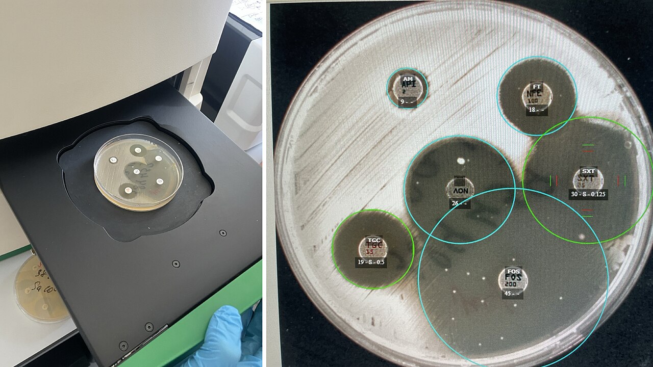
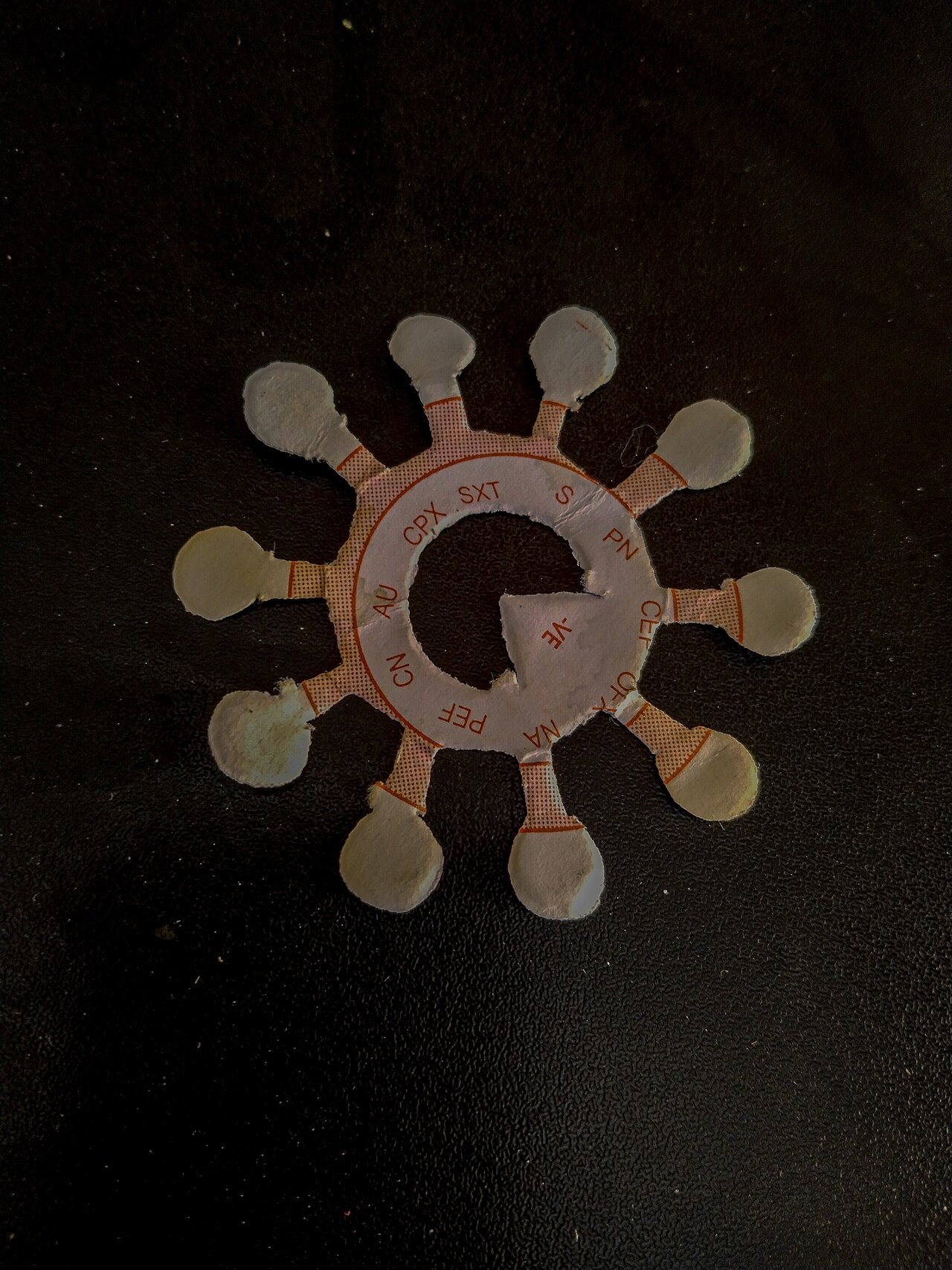
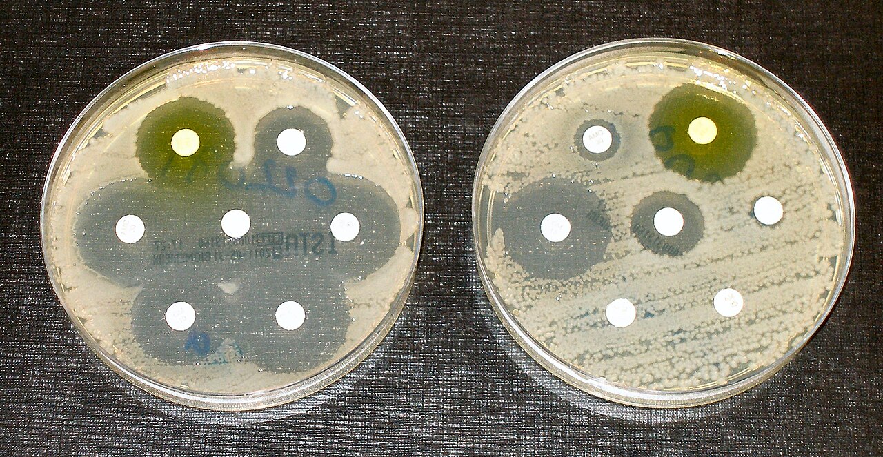
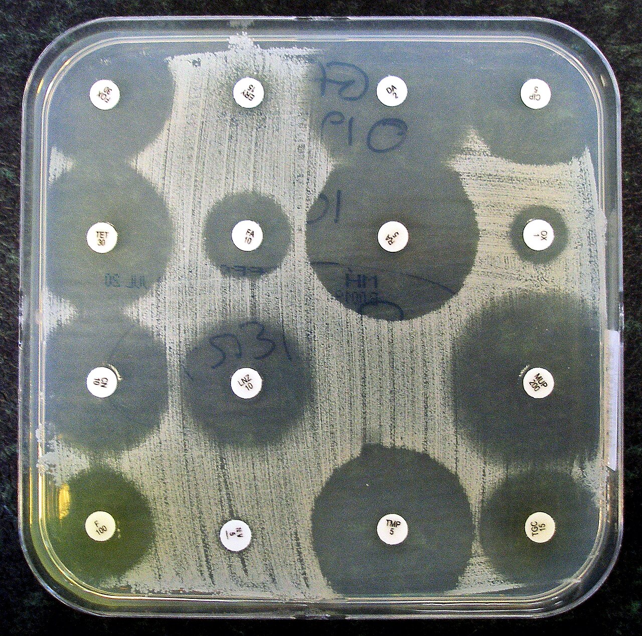
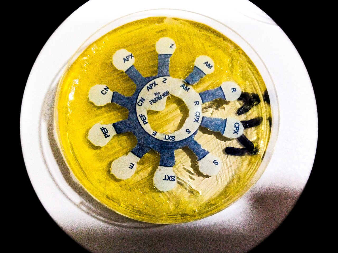
At its core, susceptibility testing asks a simple question: does this antibiotic stop this bacterium from growing? The traditional phenotypic route answers by direct observation. In the disc diffusion technique, also known as the Kirby-Bauer method, a standardized bacterial lawn is spread across an agar plate, and small paper discs soaked in specific antibiotics are laid on the surface. If the drug is effective, a clear circle of no-growth—called a zone of inhibition—appears around the disc. The diameter of that ring is compared against reference thresholds to classify the organism as sensitive, intermediate, or resistant, and to estimate the minimum inhibitory concentration. A related approach, the Etest, uses an antibiotic-impregnated strip rather than a disc and has been in clinical use since the 1980s. On the genetic side, polymerase chain reaction assays, available since the early 2000s, detect specific resistance-conferring genes directly in the bacterial genome, bypassing the need to grow the organism. Results can be qualitative—simply present or absent—or quantitative, expressed as an MIC value.
A Century of Refinement: From Penicillin to Microfluidics
The need to determine which antibiotics actually work against a given pathogen dates back to the very discovery of penicillin, the first beta-lactam antibiotic. In those early days, testing was almost exclusively phenotypic, relying on culture-based or dilution-based observations of bacterial growth in the presence of a drug. Over the decades, the toolkit expanded considerably. The Etest, a strip impregnated with a gradient of antibiotic, entered the laboratory in the 1980s and offered a more convenient alternative to traditional disc diffusion. The early 2000s brought a paradigm shift with the introduction of polymerase chain reaction–based genetic methods, which could identify resistance genes without ever culturing the organism. Today, the field continues to evolve. Researchers are actively working to make existing assays faster and more precise, while entirely new platforms—microfluidics among them—are being explored as next-generation testing technologies. The trajectory is clear: what began as a simple observation of bacterial growth on a petri dish is now a rapidly advancing discipline at the intersection of microbiology, genetics, and engineering.
Guarding Accuracy: Standardization and Quality Control in the Lab
Even the most well-designed susceptibility test can produce misleading results if experimental conditions drift. Factors such as instrument malfunction, ambient temperature fluctuations, moisture levels, and the actual potency of the antimicrobial agent can all shift outcomes. To counter these risks, laboratories perform routine quality control testing using reference bacterial strains whose resistance profiles are well documented. Organizations like the American Type Culture Collection and the National Collection of Type Cultures supply these certified strains, allowing labs to verify that their equipment and reagents are performing within expected ranges. Standardization extends to every step of the procedure. Before bacteria are spread onto Mueller-Hinton agar, the inoculum must be adjusted to a precise density, typically the 0.5 McFarland standard, achieved by visually matching turbidity against reference solutions or by photometric measurement. Bodies such as the Clinical and Laboratory Standards Institute and the European Committee on Antimicrobial Susceptibility Testing publish detailed specifications governing agar type and depth, incubation temperature, and the analytical criteria used to interpret zone diameters. Together, these layers of standardization ensure that a result generated in one laboratory is comparable to one generated in another.
Gallery

More in Microbiology 1-24
Elsewhere in the Microbiology universe
Spotted an error? Know more?
This is a living reference — every entry is fact-audited, and reader corrections feed straight into our audit queue. Suggest an edit · See this site's audit record