
Amoebozoa
A major group of amoeboid protists, sister to fungi and animals.

148LENIN · CC BY-SA 4.0
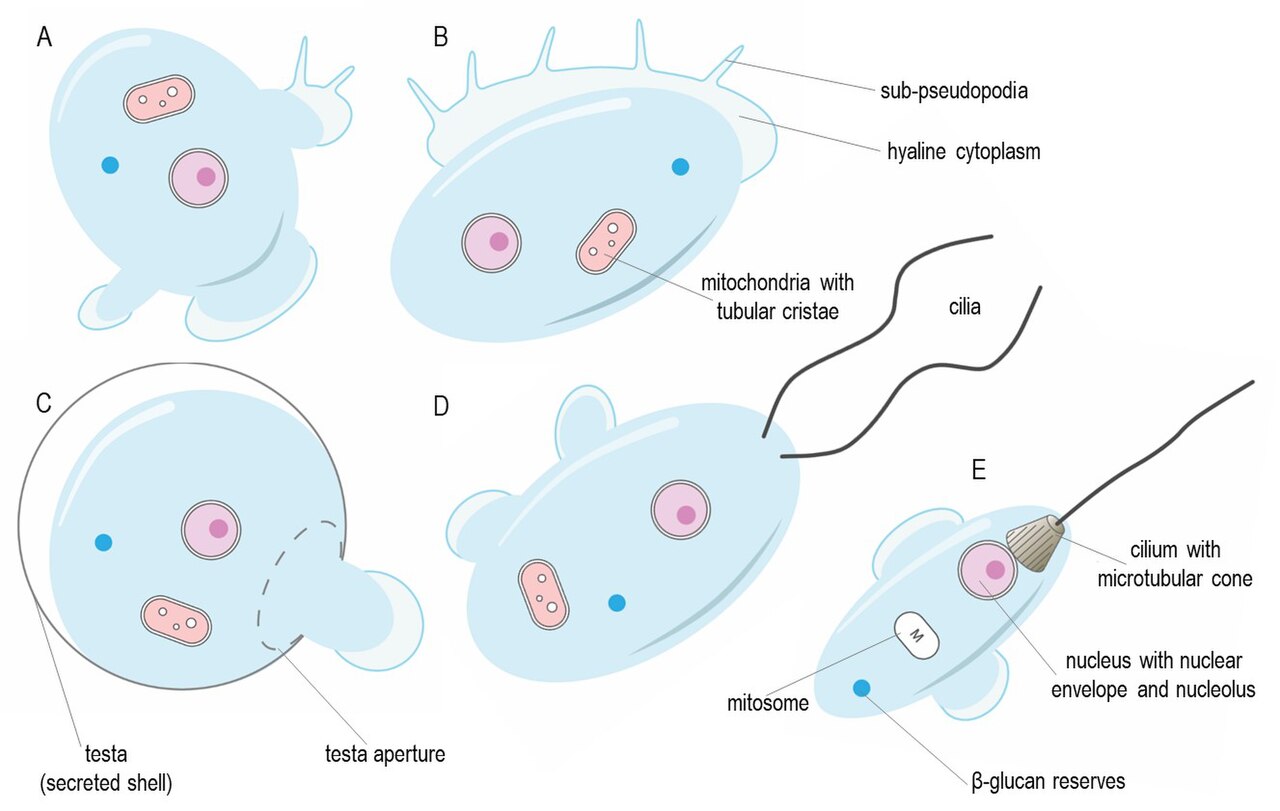
They are characterized by blunt, fingerlike, lobose pseudopods and tubular mitochondrial cristae. In traditional classification schemes, Amoebozoa is usually ranked as a phylum within either the kingdom Protista or the kingdom Protozoa, while the International Society of Protistologists retains it as an unranked supergroup within Eukaryota. Molecular genetic analysis supports Amoebozoa as a monophyletic clade, and modern studies identify it as the sister group to Opisthokonta, which includes fungi and animals.
- sister_group
- Opisthokonta
- higher_taxon
- Amorphea (with Opisthokonta)
- typical_size_range
- 10–20 μm to several square meters (multicellular forms)
- notable_genera
- Chaos, Entamoeba, Pelomyxa, Amoeba
- habitat
- salt and freshwater, soil, moss, leaf litter; some parasitic or symbiotic
Lore & Background
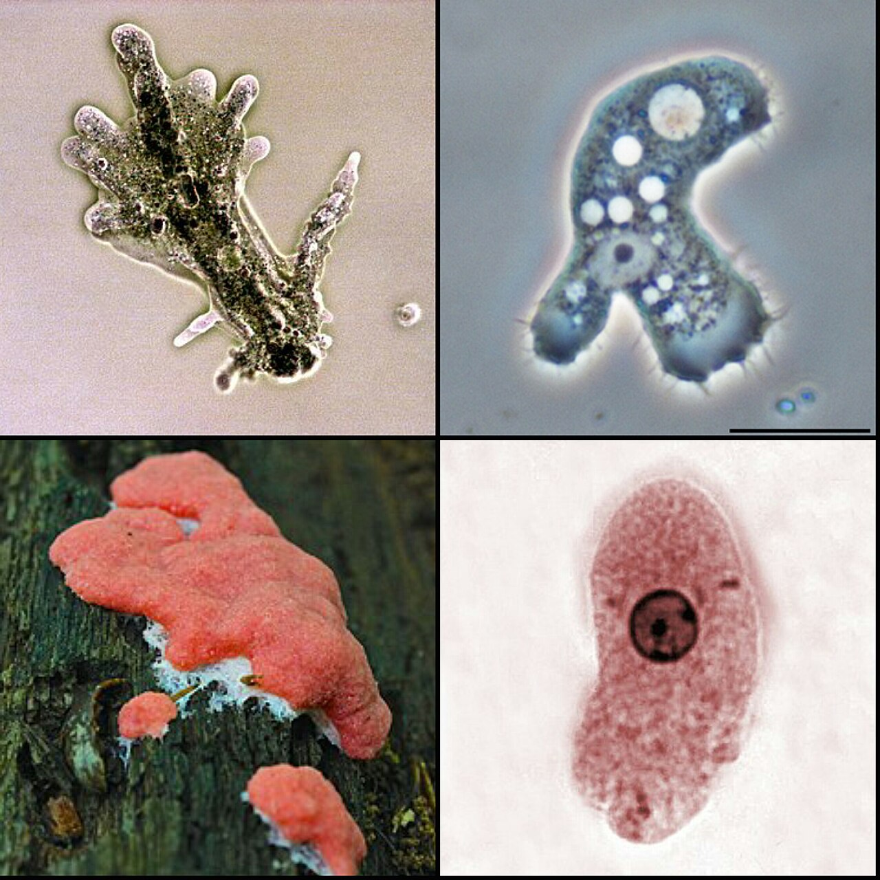
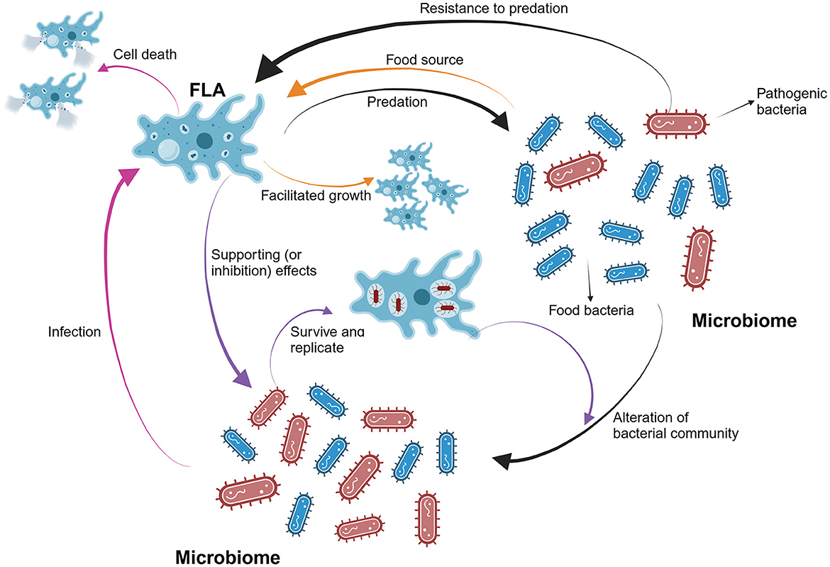
Amoebozoa includes many of the best-known amoeboid organisms, such as Chaos, Entamoeba, Pelomyxa, and the genus Amoeba itself. Species may be shelled (testate) or naked, and cells may possess flagella. Free-living species are common in both salt and freshwater as well as soil, moss, and leaf litter. Some live as parasites or symbionts, and some cause disease in humans and other organisms. While the majority are unicellular, the group also includes several clades of slime molds, which have a macroscopic, multicellular stage of life, forming a plasmodium or aggregating to form one.
Reader's Guide
Amoebozoa is significant as a major lineage of eukaryotes, forming the sister group to Opisthokonta (which includes fungi and animals). This relationship, supported by molecular genetic analysis, places Amoebozoa at a key position for understanding the early evolution of eukaryotes. The group's diversity—from unicellular amoebae to macroscopic slime molds—illustrates a range of life strategies, including phagocytosis, cyst formation, and multicellular aggregation. Amoebozoa also includes model organisms like Amoeba proteus, studied in schools and laboratories for its convenient size. The classification of Amoebozoa has been refined through structural and genetic studies, with recent work supporting a primary division into Lobosa and Conosa. The group's position in the eukaryotic tree, alongside Opisthokonta in the clade Amorphea, highlights its evolutionary importance.
Did You Know?
- Some multicellular slime molds, like Fuligo septica, can cover an area of several square meters.
- Amoebozoa and Opisthokonta are sometimes grouped together in a high-level taxon named Amorphea.
Evolutionary Position and Taxonomic Identity
For much of the twentieth century, taxonomists slotted this group into the kingdom Protista or Protozoa as a phylum, but the International Society of Protistologists now treats it as an unranked supergroup nested within Eukaryota. Because of this tight pairing, researchers have proposed umbrella names for the combined group. Thomas Cavalier-Smith originally coined Unikonta, envisioning a common ancestor bearing a single emergent flagellum, but subsequent work has undermined that uniciliate hypothesis.
Cell Architecture and the Mechanics of Movement
A typical amoebozoan cell is internally partitioned into two functional zones: a granular core known as endoplasm and a transparent peripheral layer called ectoplasm. During active locomotion, the endoplasm surges forward while the ectoplasm streams backward along the cell's outer surface, creating a continuous circulatory flow. Many species adopt what is termed a monopodial posture, in which the whole body effectively acts as one large pseudopod. From that leading pseudopod, organisms can extend numerous fine, clear projections called subpseudopodia or determinate pseudopodia; these reach a set length before retracting, serving either to pull the cell forward or to capture prey. Alternatively, a cell may deploy several indeterminate pseudopodia, tubular extensions packed with granular endoplasm, into which the entire cell mass flows. The leading pseudopod advances while trailing ones retract, unless the organism simply changes heading. Externally, most members are naked like the familiar Amoeba or Chaos, some wear a loose coat of tiny scales as seen in Cochliopodium and Korotnevella, and members of the order Arcellinida build rigid shells called tests, either secreted from organic material as in Arcella or assembled from cemented environmental particles as in Difflugia, with a single aperture through which pseudopods emerge.
Ecological Breadth and the Slime Mold Connection
Amoebozoa occupy an astonishing range of habitats and ecological niches. Free-living species thrive in salt water, freshwater, soil, moss, and leaf litter, while others exist as parasites or symbionts of other organisms, and a subset is known to cause disease in humans and other hosts. The group is dominated by unicellular organisms, yet it also harbors several clades of slime molds that undergo a striking multicellular phase. In plasmodial slime molds, individual amoeboid cells continue dividing while remaining physically joined, producing a macroscopic plasmodium. Cellular slime molds take a different route: separate cells aggregate into a coordinated multicellular structure. Size variation within the group is extreme. Multinucleate genera such as Chaos and Pelomyxa reach several millimeters, and the dog vomit slime mold Fuligo septica can blanket areas spanning several square meters. Mixotrophic species in Arcellinida and Mayorella even host microalgae of the genus Chlorella within their own cytoplasm, blurring the boundary between animal and plant modes of nutrition.
Feeding, Survival Structures, and Cellular Machinery
Every amoebozoan feeds by phagocytosis: pseudopods extend around a food particle, seal it into an intracellular vacuole, and the contents are then digested and absorbed. Some species possess a posterior bulb called a uroid that collects metabolic waste and periodically pinches off from the main body. When conditions turn harsh and food becomes scarce, most amoebozoans encyst; these dormant shells can be carried on air currents to entirely new environments. In slime molds, the analogous survival structures are spores borne on stalked fruiting bodies or sporangia. At the cellular level, the majority of the group lacks flagella and does not build microtubule-supported structures outside of mitosis. Exceptions do exist: Archamoebae bear flagella, and many slime molds produce biflagellate gametes. The flagellum in these cases is anchored by a cone of microtubules, a feature that echoes the opisthokont lineage and reinforces the close evolutionary tie between the two clades. Mitochondria across Amoebozoa characteristically display branching, tubular cristae, though Archamoebae adapted to anoxic or microaerophilic niches have lost their mitochondria entirely.
Gallery
More in Microbiology 1-24
Elsewhere in the Microbiology universe
Spotted an error? Know more?
This is a living reference — every entry is fact-audited, and reader corrections feed straight into our audit queue. Suggest an edit · See this site's audit record