Bacterial cell structure
Bacteria have unique cell structures including peptidoglycan cell walls.

Shahid Khan · CC BY-SA 4.0
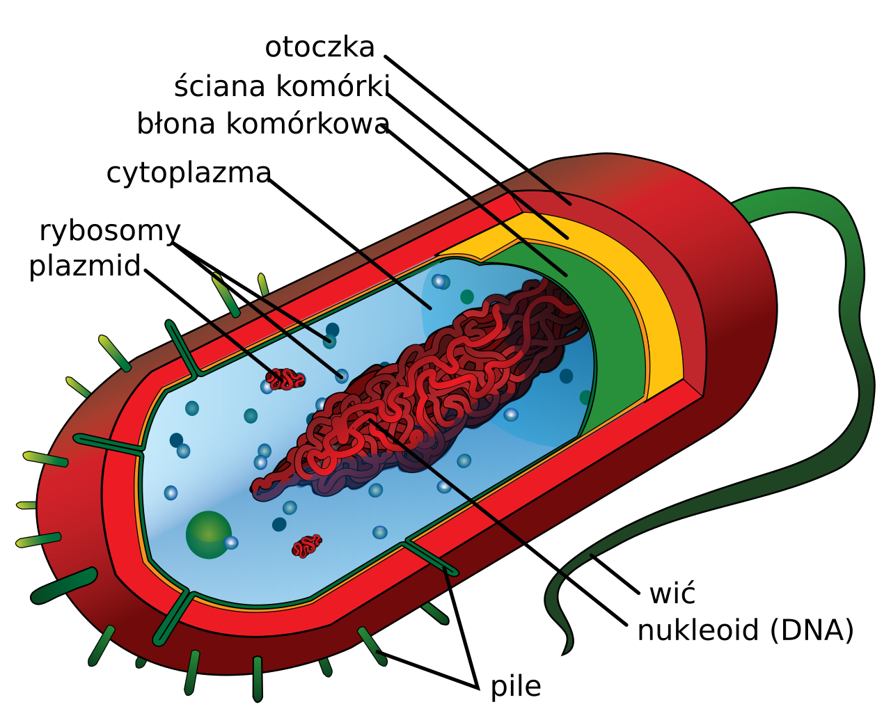
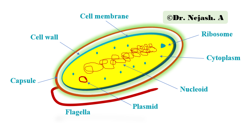
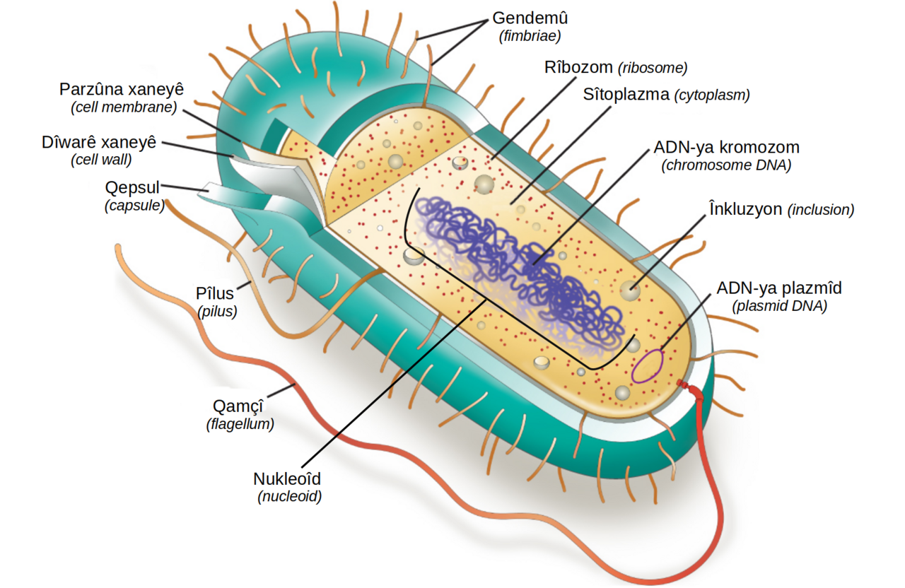
Bacteria are prokaryotic microorganisms with highly organized and efficient cell structures that allow them to occupy diverse ecological niches. Their cells have fewer components than eukaryotic cells, making them easier to manipulate experimentally, and many biochemical principles were first characterized in bacteria before being applied to other organisms.
- cell_size
- typically 0.5–2 μm
- cell_volume
- 0.6–0.7 μm³ (E. coli)
- dry_mass
- about 0.2 pg per cell
- cell_wall_component
- peptidoglycan
- gram_types
- gram-positive and gram-negative
Lore & Background
Bacterial cells exhibit diverse morphologies such as coccus, bacillus, coccobacillus, spiral, and filamentous forms, which are traditionally important for identification and classification. Cell shape is generally characteristic of a given species but can vary with growth conditions. Some bacteria, like Caulobacter, produce stalks and appendages, while others, like Myxococcus and Streptomyces, form elaborate structures bearing reproductive spores. The cell envelope consists of the cell membrane and the cell wall, with peptidoglycan providing structural integrity and determining cell shape. Peptidoglycan is made of alternating N-acetylmuramic acid and N-acetylglucosamine residues. Gram-positive bacteria have thick peptidoglycan layers (up to 95% of the wall) and stain purple, while gram-negative bacteria have thinner peptidoglycan and an additional outer membrane containing lipopolysaccharides, staining pink. Gram-positive walls often contain teichoic acids and lipoteichoic acids, which anchor peptidoglycan to the membrane and confer a negative charge. Outside the wall, many gram-positive bacteria have an S-layer of proteins and a polysaccharide capsule. Gram-negative walls have porins in the outer membrane for passive transport, and the periplasm between membranes contains the peptidoglycan layer and various proteins.
Reader's Guide
Bacterial cell structure is fundamental to microbiology because it underpins the survival, classification, and pathogenicity of bacteria. The presence of peptidoglycan in the cell wall is unique to bacteria and distinguishes them from archaea and eukaryotes. This structural feature is the target of many antibiotics, such as penicillins and cephalosporins, which inhibit cell wall synthesis without affecting human cells. The Gram staining method, which differentiates bacteria based on cell wall composition, remains a key diagnostic tool. The small size of bacteria, with a high surface area-to-volume ratio, enables rapid nutrient uptake and waste excretion, contributing to their evolutionary fitness. Understanding bacterial cell structure has allowed the development of experimental systems that revealed fundamental biochemical principles, later applied to higher organisms. The variability in cell wall architecture between gram-positive and gram-negative bacteria influences their susceptibility to antibiotics and host immune responses, making this knowledge critical for medicine and biotechnology.
Did You Know?
- Bacterial cell walls contain peptidoglycan, which is not found in archaea or eukaryotes.
- Gram-positive bacteria have a thick peptidoglycan layer that stains purple, while gram-negative bacteria have a thinner layer and an outer membrane, staining pink.
- The enzyme lysozyme, found in human tears, digests bacterial cell walls and is the body's main defense against eye infections.
- Teichoic acids are found only in gram-positive bacteria and give the cell wall an overall negative charge.
Shape, Size, and the Geometry of Survival
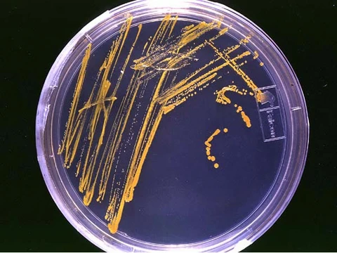
Bacteria come in a striking variety of body plans—round cocci, rod-shaped bacilli, intermediate coccobacilli, corkscrew spirals, and elongated filaments—that have historically anchored their identification and classification. A species typically holds a characteristic shape, yet growth conditions can nudge it. A handful of lineages add further flair: Caulobacter extends stalks and appendages through its life cycle, while Myxococcus and Streptomyces construct elaborate spore-bearing structures. Under light microscopy these individual forms become visible, and on a Petri plate they resolve into distinctive colony patterns. Their smallness is equally defining. An Escherichia coli cell spans roughly two micrometres in length and half a micrometre in width, enclosing about 0.6 to 0.7 cubic micrometres and carrying a wet mass near one picogram. Dry mass is around 0.2 picograms, split roughly evenly between carbon and protein. This compact geometry is no accident: a high surface-area-to-volume ratio accelerates nutrient uptake and waste excretion, keeping metabolism swift. When the ratio falls, diffusion across the membrane becomes rate-limiting and the cell's fitness declines. Why some bacteria grow larger remains uncertain, though the extra volume may simply serve as a storage depot for surplus nutrients.
Peptidoglycan: The Architectural Signature of Bacteria
The bacterial cell envelope pairs a cell membrane with a cell wall, and the wall's chief job is to shield the cell from the internal turgor pressure generated by the high concentration of proteins and other solutes inside. What sets bacterial walls apart from every other organism's is the presence of peptidoglycan, a polymer laid down immediately outside the membrane. Its polysaccharide backbone alternates N-acetylmuramic acid and N-acetylglucosamine residues in equal numbers, and this meshwork is responsible for both the rigidity of the wall and the determination of cell shape. Despite its structural role, peptidoglycan is fairly porous; particles on the order of two nanometres can pass through it, so it does not act as a permeability barrier for small substrates. With rare exceptions such as the extracellular parasite Mycoplasma, every bacterial species builds peptidoglycan into its wall, yet the overall architecture varies widely. If the wall is stripped away entirely the cell is termed a protoplast; if only partially removed, a spheroplast.
Gram Staining, Teichoic Acids, and the Antibiotic Frontier
Bacterial cell walls fall into two broad categories—gram-positive and gram-negative—distinguished by how they take up crystal violet during Gram staining. In gram-positive organisms the peptidoglycan layer is thick, constituting up to 95 percent of the wall in some species, whereas in gram-negative bacteria it accounts for only five to ten percent. The Deinococcota group is a curious hybrid: it stains gram-positive yet harbours some wall structures typical of gram-negative cells. Beyond peptidoglycan, gram-positive walls often contain teichoic acids, polymers of either ribitol phosphate or glycerol phosphate found exclusively in this group. Glycerol teichoic acids are the more widespread form, though their precise biological role remains debated. Some are lipid-linked, forming lipoteichoic acids covalently attached to lipids within the cytoplasmic membrane. The wall's vulnerability underpins a therapeutic strategy. Beta-lactam antibiotics such as penicillin and cephalosporin block the cross-linking of peptidoglycan, halting wall synthesis and killing the bacterium without harming human cells, which lack a wall. The enzyme lysozyme, found in human tears, digests the bonds between NAM and NAG residues and serves as the body's main defence against eye infections. Some gram-positive species, notably Staphylococcus aureus, resist lysozyme by adding O-acetyl groups to certain muramic acid residues.
Bacteria as the Workbench of Biochemistry
Although they look deceptively simple, bacterial cells are highly organized and remarkably efficient, a fact that has allowed them to colonise an enormous range of ecological niches. They share a prokaryotic body plan with archaea, yet many structural features are unique to bacteria and absent from both archaea and eukaryotes. This distinctiveness, combined with a smaller inventory of cellular components than eukaryotic cells, makes bacteria far easier to manipulate experimentally. As a result, a great many biochemical principles were first worked out in bacterial systems before researchers could translate those findings to more complex organisms. The relative simplicity of the bacterial cell—fewer components, a straightforward envelope—has made it an ideal reductionist platform for probing fundamental biology. From the initial characterisation of biochemical principles to their subsequent application across the tree of life, bacteria have served as the foundational model on which much of modern biology was built. Their study continues to underpin advances in the life sciences, a legacy that flows directly from the very structural economy that makes them so small, so fast, and so easy to work with.
Gallery
More in Microbiology 1-24
Elsewhere in the Microbiology universe
Spotted an error? Know more?
This is a living reference — every entry is fact-audited, and reader corrections feed straight into our audit queue. Suggest an edit · See this site's audit record